AVIAN/FUR/EXOTIC SPECIES
Rabbit hepatic coccidiosis- re-emerging as a clinical disease?
Marina Brash
Liver samples were submitted to the AHL for histologic evaluation from a postmortem conducted on a rabbit that was one of multiple rabbits that had died suddenly with no evidence of clinical illness. This rabbit was one of many that were housed in a large pen on a small urban farm. The only gross lesion that was identified on postmortem was a liver filled with tiny abscesses (Fig. 1) and histologically the abscesses were determined to be dilated and fibrotic bile ducts containing large numbers of coccidial organisms (Figs. 2, 3). Wet-mount examination of fluid from these cystic nodules is a useful in-clinic test that quickly demonstrates the characteristic ovoid Eimeria stiedae oocysts measuring ~30 X 20 µm (Fig. 4). Demonstration of coccidial oocysts by fecal flotation however, is not diagnostic, for the rabbits are also likely shedding intestinal coccidial oocysts that are of a mixture of species.
Enteric coccidiosis continues to be one of the important potential causes of enteritis in Ontario commercial rabbitries, but hepatic coccidiosis has seldom been identified in rabbits submitted to the AHL for diagnostic enteritis workup. However, over the last few years, there has been increased interest in the raising of small groups of rabbits for meat or as pets and these rabbits have either been raised on the ground or allowed access to the ground. Owners of these small rabbitries have contacted their veterinarians for morbidity and mortality concerns and in turn, the AHL has received increased enquiries and pathology submissions from veterinarians related to the identification of hepatic coccidiosis in these rabbits.
In commercial rabbitries, hepatic coccidiosis continues to be a concern but typically occurs as a sub-clinical disease resulting in condemnations of livers from meat-type rabbits at processing. However, if young, naive rabbits are exposed to high enough levels of sporulated oocysts, clinical disease including anorexia, poor weight gain, distended abdomens, diarrhea, and death can occur. Long-lasting immunity to Eimeria stiedae is possible, if the rabbits do not receive an excessively high exposure initially and are immunocompetent.
Coccidial oocysts are extremely resistant to environmental influences and no commonly available disinfectants will kill coccidial oocysts, so minimizing the exposure to infected feces is of great importance. This will be challenging if the rabbits are raised or are allowed access to the ground and the concern is that hepatic coccidiosis may be re-emerging as a clinical disease. AHL
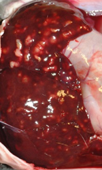
Figure 1. Characteristic appearance of hepatic coccidiosis caused by Eimeria stiedae. The liver is enlarged with numerous raised, firm, pale, variably sized, cystic, round to elongate nodules filled with turbid pale green-yellow fluid scattered throughout the hepatic parenchyma.
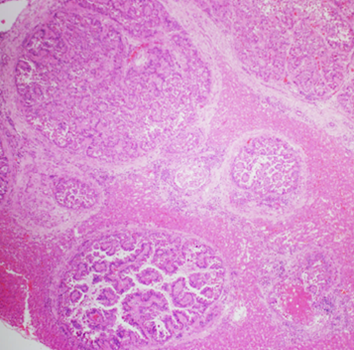
Figure 2. The liver nodules described by the veterinarian were dilated bile ducts lined by hyperplastic biliary epithelium rimmed with increased amounts of periductal fibrous tissue with compression of the surrounding hepatic parenchyma. 40X H&E.
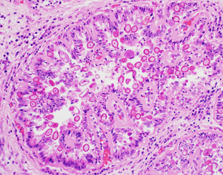
Figure 3. A dilated bile duct. Biliary epithelial cells are filled with asexual and sexual developmental stages of coccidial organisms and the lumen contains coccidial oocysts. 200X H&E.
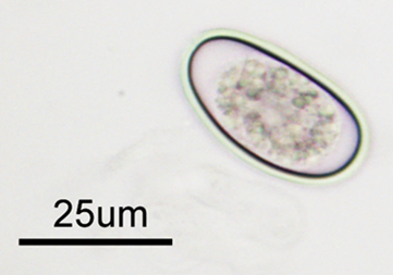
Figure 4. The characteristic ovoid Eimeria stiedae oocysts measuring ~30 X 20 µm can be demonstrated in wet mount preparations made from the fluid aspirated from the cystic liver nodules.