AHL Newsletter June 2018
Click here for a pdf copy of the June 2018 AHL Newsletter.
May 1, 2018, AHL User’s Guide and Fee Schedule
- Includes more test information, new tests, new test panels, fee adjustments, and more!
- Mobile friendly!
- Available on-line at https://www.uoguelph.ca/ahl/
- Test information is linked to LabNotes to facilitate test selection and interpretation of results.
- Please note that tumor margin evaluation, histt, is charged in addition to the histopathology fee (see below).
- New tests - lipid profile; insulin (RIA) and glucose; Mycoplasma hyopneumoniae - gene sequencing typing; Trichomonas gallinae - PCR; scrapie resistance PrP genotyping in deer (codon 96) - sequencing; Myxobolus cerebralis (whirling disease pathogen) - PCR; Bacillus anthracis (anthrax) - real-time PCR; Mycoplasma suis, M. wenyonii, M. ovis, M. haemolamae PCR; bromethalin (desmethylbromethalin) - LC-MS/MS; selenium, blood - ICP-MS; bluetongue virus/epizootic hemorrhagic disease virus - PCR; bovine abortion panel - PCR (BoHV-1/IBR, Leptospira, Neospora caninum); Salmonella Dublin - antibody ELISA; Leptospira spp - PCR; bluetongue virus Ab - ELISA; Sporothrix schenckii - RT-PCR. AHL
Companion animal histopathology tests at the AHL
Josepha DeLay
AHL histopathology tests and associated fees for companion animal biopsies and in-clinic postmortems are categorized based on the number and size of samples. Please be sure to read the details for each subset of test so that you select the correct test for each case. Also please note that for surgical margin evaluation of biopsies >2 cm diameter, the ‘tumor margin evaluation’ test must be selected in addition to the appropriate histopathology test. An additional fee applies for margin evaluation on these larger biopsies. Please refer to the AHL website for current fees. AHL
|
Test name |
Test code |
Details |
|
Histopathology, 1-2 biopsies or tissues |
histcm1
|
For submissions with 1-2 biopsies or tissues, OR multiple (6 or fewer) punch, trucut, or endoscopic biopsies. |
|
Histopathology, 3-6 biopsies or tissues |
histcm2
|
For submissions with 3-6 biopsies or tissues, OR biopsies 6-10 cm diameter. |
|
Histopathology, 7 or more biopsies or tissues |
histcm3
|
For submissions with 7 or more biopsies or tissues, OR biopsies / samples >10 cm diameter (eg large tumors, spleen, brain, mammary chain, heart). |
|
Tumor margin evaluation |
histt
|
Applies in addition to regular histopathology charge. For tumor excisional biopsies >2 cm diameter. Includes preparation of 4 radial margin sections. Must be requested at time of sample submission. |
Client Outreach Technician - Josie Given
Jim Fairles
The AHL is pleased to announce that, under the Ontario Animal Health Network, Josie Given has been named to the permanent position of Client Outreach Technician - https://www.uoguelph.ca/ahl/people/josie-given .
Josie will continue to focus on the voluntary AHL in-clinic milk bacteriology quality program
https://www.uoguelph.ca/ahl/content/ahl-labnote-47-ahl-milk-bacteriology-clinic-laboratory-proficiency-program as well other areas of the in-clinic laboratory. She will also concentrate on helping clinics get the most from their submissions to the lab. This will include demonstration and implementation of the new AHL Client Portal. This portal will provide a user-friendly online submission process in which clinic client information can be stored and retrieved easily. This will aid in giving the AHL the best information needed for testing and scanning surveillance including demographics, premises identification numbers (PIDs), and history.
Josie brings a wealth of experience from her experience in this position under the Disease Surveillance Plan. Please join us in welcoming Josie to this position. AHL
Small Ruminant Adult Mortality Project deadline approaching - only 2 months left to submit cases!
Maria Spinato
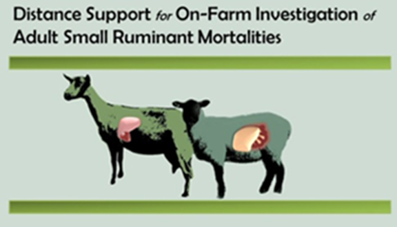
- Last date to submit a case is July 31, 2018.
- 89 cases completed to date.
- Sufficient funds remain to test 80 more cases.
- Postmortems and submitting samples during the warmer months: if the tissues are “green”, the case will be rejected as bacterial cultures will be overgrown and histopathology will be non-diagnostic
- If the samples cannot be shipped overnight or dropped off at AHL, fresh tissue samples and head should be frozen.
- Please check your collection kit to ensure that the formalin and culture swabs are not expired.
AHL Newsletter
June, 2018 - Volume 22, Number 2
Editor: Grant Maxie, DVM, PhD, Diplomate ACVP
Editorial Assistants: Helen Oliver, April Nejedly
The AHL Newsletter is published quarterly (March, June, September, December) by the Animal Health Laboratory, Laboratory Services Division, University of Guelph.
Its mission is to inform AHL clients and partners about AHL current activities, and laboratory-based animal disease events and disease trends. All material is copyright 2018. Ideas and opinions expressed herein do not necessarily reflect the opinions of the University or the Editor.
Articles may be reprinted with the permission of the editor and with appropriate credit given to the AHL Newsletter.
Mailing address & contact information:
Animal Health Laboratory
Laboratory Services Division, University of Guelph
Box 3612, Guelph, Ontario, Canada N1H 6R8
Phone: (519) 824-4120 ext. 54538; fax: (519) 821-8072
To receive an electronic copy of this Newsletter, please send your email address to us at holiver@uoguelph.ca
ISSN 1481-7179
Canada Post Publications number - 40064673
Contributors to this issue
- from the Animal Health Laboratory:
Melanie Barham, DVM, PMP
Marina Brash, DVM, DVSc, Diplomate ACVP
Andrew Brooks, DVM, PhD, Diplomate ACVP
Michael Deane, BA
Josepha DeLay, DVM, DVSc, Diplomate ACVP
Jason Eidt, BSc
Jim Fairles, DVM, MBA
Murray Hazlett, DVM, DVSc, Diplomate ACVP
Beverly McEwen, DVM, PhD, Diplomate ACVP
Felipe Reggeti, DVM, PhD, Diplomate ACVP
Janet Shapiro, DVM, DipEqSurg, DipPath
Durda Slavic, DVM, PhD
Maria Spinato, DVM, DVSc, Diplomate ACVP
Quimei You, DVM, MSc
Jen Zoethout, BScH
- Other contributors:
Al Dam, BSc; Csaba Varga, DVM, PhD, OMAFRA, Guelph
John Hancock, DVM, Picton, ON
Wilson Rumbeiha, BVM, PhD, DABT; Dwayne Schrunk, BS, Veterinary Diagnostic Lab, Iowa State University, Ames Iowa
Sherry Smith, DVM, Dunnville, ON
Kathleen Taylor BSc, DVM; Bruce Watt DVM, Dipl ACVS, Uxbridge, ON
Lloyd Weber, DVM, Guelph, ON
Our continued thanks to all of the non-author AHL clerical, technical, and professional staff who contribute to the generation of results reported in the AHL Newsletter.
Ontario Animal Health Network
OAHN Update, June 2018

OAHN project videos and reports: In 2015, each network was funded to fill a gap in surveillance through a project. OAHN projects were run by a Principal Investigator in conjunction with each network. You can watch videos we have filmed with the grad students who were working on OAHN projects here: http://oahn.ca/news/oahn-grad-student-features/.
OAHN program announcement: As of May 1st, 2018, OAHN will continue within the OMAFRA-University of Guelph agreement, and will continue to be housed at AHL, in collaboration with OMAFRA, UofG, and private industry. We are ecstatic that this project has become a program, and are looking forward to continuing with Ontario animal disease surveillance and spreading the word to veterinarians, producers, owners, and others. To read about what we accomplished in our first 5 years, check out the DSP Magazine: https://oahn.ca/resources/staying-ahead-of-the-curve-magazine-the-disease-surveillance-plan-2013-2018/
Recent news featuring OAHN: OAHN was featured in Research Magazine, published by the OMAFRA-University of Guelph Agreement. Check us out on pp 20-21: https://www.uoguelph.ca/research/sites/default/files/public/UofG_Research_Yearbook_2017-18_Web_sm.pdf
New look for OAHN reports: Stay tuned for our new look for both producer and veterinary reports this quarter.
 New Podcasts: Ticks and Rabies in Ontario
· 2018 Ontario Rabies Update http://oahn.podbean.com/e/2018-ontario-rabies-update-with-dr-maureen-anderson/
· Ontario Tick-Borne Disease Update http://oahn.podbean.com/e/ontario-tick-borne-disease-update-with-dr-michelle-evason/
Staying Up-to-Date on Ontario Animal Health
We regularly publish disease alerts and industry updates that are sent to us from OMAFRA, the Feather Board Command Centre, Public Health Units, and other reputable Ontario organizations. In addition, we scan the internet, publishing current animal health and welfare, One Health, and veterinary medicine news from Ontario and throughout North America. There are 4 ways you can follow us to stay on top of animal health news:
· OAHN Facebook Page—https://www.facebook.com/OntarioAnimalHealthNetwork/
· OAHN Twitter Page—https://twitter.com/OntAnHealthNet
· Follow the OAHN News Section—https://oahn.ca/news-archive/
· Sign-up for the OAHN Veterinary Newsletter if you’re a vet or RVT.
New Infographics
The OAHN Equine Network produced 2 infographics on West Nile virus for equine veterinarians to share with their clients, promoting the value of vaccination: http://oahn.ca/resources/oahn-equine-west-nile-virus-infographics/
*New Network Reports*
We have recently published new veterinary and/or owner/producer reports for our Poultry, Companion Animal, Small Ruminant, Swine, and Equine networks. You can access the reports by going to https://oahn.ca and clicking on the Networks Reports tab. Note that you must be registered and signed in to view the veterinary reports, along with lab data and clinical impression summaries.
Do you ask your clients for their Premises IDs? Check out how fast and easy it is to do by registering your vet clinic: https://www.ontarioppr.com/home_en.html
Questions and LabNote 50
Why can’t I use a Ziploc for submissions? (and other excellent questions about submission containers)
Melanie Barham, Jim Fairles
 We love receiving samples in the best possible condition, as it truly helps to improve our ability to provide our clients with useful answers. Below, we’ve addressed some of the most common packaging errors seen:
Why do I need to use a Whirl-Pak bag? Won’t a zipper sandwich bag do?
Whirl-Paks are preferred because they are sterile inside, which is particularly important if submitting tissue for bacteriology culture. Whirl-Paks also don’t leak in the same way zippered sandwich bags do. Although they may leave your clinic in optimal condition, often samples packed in sandwich bags leak in transit. Whirl-Paks also comply with Transportation of Dangerous Goods regulations. Have new staff who have never used Whirl-Paks? Check out our handy video: https://www.youtube.com/watch?v=27oT0BUHOr0
How should I submit my fecal samples?
Although we all love surprises, being surprised with a fecal matter explosion when opening a box of samples is not as much fun as it might sound. We appreciate receiving samples in screw-top containers. Red-top tubes often pop their lids due to gas-producing microbes, and gloves with feces in the fingers takes a significant amount of excess processing time (delaying your results), and they often burst or leak. We are pleased to provide fecal containers for purchase, although sterile urine containers, mastitis sampling jars, or other sterile plastic screw top containers are also acceptable.
What samples frequently arrive broken or in poor quality?
Glass vials! Blood tubes often arrive broken or cracked. A simple fix to this is to wrap a piece of newsprint, paper towel, or bubble wrap between vials.
Formalin jars! Formalin jars arrive leaking quite frequently. This can be a problem for the individuals couriering the samples, and often delays your samples as submission forms can become illegible if liquid leaks on them. A simple fix is to seal jars tightly, wrap in Parafilm or tape, and wrap in bubble wrap.
Submission forms! Submission forms should be packaged separately from samples in case of leakage. Consider using the outside pocket of the laboratory zipper seal bag (it’s not just there for decoration!), or placing the submission form in a different zipper seal bag for multiple samples. We also offer online submission forms that can be filled out from any mobile device or desktop to save you paperwork. AHL
LabNote 50 - Postmortem submissions via courier
Jim Fairles, Jen Zoethout
AHL realizes that, because of distance, not everyone can drive to our labs in Guelph or Kemptville to drop off bodies for postmortem. AHL will accept bodies for postmortem via courier – precautions need to be taken.
Please see our submission instructions: http://www.guelphlabservices.com/ahl/submissions/ahl-labnote-27-submission-instructions
Postmortem submissions also need to abide by the following
* Use AHL’s Purolator return waybill – “Animal Specimen Exempt” should be on the label.
* Pack in an impervious container (e.g., Rubbermaid container and triple bag with cold packs). Leaks must be avoided.
* Call 519-824-4120 ext 54530 or email (specroom@uoguelph.ca) AHL with submission tracking number to give heads-up that samples are arriving.
* Ship Monday to Wednesday for next-day delivery to avoid weekend delays.
Because of the weight, courier costs for these shipments are considerably higher than normal sample packages delivered to AHL.
Food animal courier costs will be waived. However, we will charge back some of the courier cost for companion animal shipments over 5 kg. $25 per each 20 kg - new test code xtrnpm. This charge will be added to the invoice for the case. AHL
RUMINANTS
Mortality in lambs caused by Fascioloides magna
Andrew Brooks, Jan Shapiro, John Hancock
Several lambs were submitted to AHL-Kemptville for postmortem examination with a history of increased mortality and sudden death affecting a group of ram lambs ~ 10 mo of age from a farm in eastern Ontario.
Postmortem findings included numerous regions of cavitation and necrosis in the livers (Fig. 1), with abscesses containing dark brown exudate, and chronic adhesions of the liver to the adjacent viscera and diaphragm. Some lambs had abscesses in the peritoneum and pelvis, and black pigment in the omentum and liver (Fig. 2). Many lambs also had pneumonia, fibrotic pleural adhesions, acute pulmonary hemorrhage (Fig. 3), and pulmonary cavities containing dark brown exudate.
Serial sectioning of the livers revealed a large fluke ~ 2 x 1.8 x 0.4 cm with morphology compatible with Fascioloides magna (Fig. 4). Sequencing of the 18S rRNA gene confirmed the identity of the parasite (99.8% sequence similarity to F. magna).
Histopathology revealed widespread necrosis and abscessation of the liver and lung, with concurrent bacterial and mycotic infections that were interpreted as secondary. Bacteria such as Pasteurella multocida, Yersinia pseudotuberculosis, and Mannheimia glucosida were isolated.
F. magna (large American liver fluke) is primarily a parasite of cervids, particularly white-tailed deer, which are the definitive hosts. In deer, the flukes reside in cysts in the liver parenchyma and eggs are eventually excreted in the feces via connections between the cysts and bile ducts. Freshwater snails such as Lymnaea spp. are the intermediate hosts. In cattle, the flukes migrate briefly in the liver and become encysted, but in sheep they migrate continuously resulting in severe damage to the liver and occasionally the lungs. Infection of sheep with only a few F. magna flukes can result in death. Sheep and cattle become infected by ingesting infective metacercariae on pastures shared with parasitized deer and freshwater snails.
Heavier than usual rainfall in eastern Ontario in 2017 created very wet pasture conditions with some flooding, which may have played a role in the transmission of the parasite that had not been diagnosed here for many years. AHL
Reference:
Taylor MA, et al. Veterinary Parasitology. Wiley Blackwell, 2016:76-77;486, 766.

Figure 1. Severe cavitation and necrosis in the liver.

Figure 2. Black pigment in the mesentery and omentum.

Figure 3. Acute hemorrhage in the lung and pleura.

Figure 4. Fascioloides magna fluke found in the liver.
RUMINANTS
Yersinia pseudotuberculosis enterocolitis and lymphadenitis in a goat
Maria Spinato, John Hancock, Durda Slavic
A 2-y-old Boer goat doe died following a short bout of diarrhea. During an on-farm postmortem, significant external findings included moderate dehydration, sunken eyes, and liquid feces soiling the hind end. The most remarkable internal lesions were depleted fat stores and marked enlargement of mesenteric lymph nodes (Fig. 1). Formalin-fixed and fresh tissue samples were submitted to the Animal Health Laboratory for testing, as per the small ruminant adult mortality project protocol.
Histologically, the most remarkable lesions included bacterial microabscesses within sections of mesenteric lymph node (Fig. 2), erosive and necrotizing enterocolitis accompanied by proliferation of large bacterial colonies (Fig. 3), and multifocal necrotizing hepatitis typified by colonies of bacterial coccobacilli surrounded by a broad rim of degenerate leukocytes. Heavy (3+) growths of Yersinia pseudotuberculosis were isolated from cultures of jejunum and a swab of mesenteric lymph node.
This doe died of Yersinia pseudotuberculosis septicemia; localizing lesions predominantly involved the small intestine (diarrhea), mesenteric lymph nodes (lymphadenopathy), and liver. This organism is a member of the gastrointestinal flora of multiple species of animals, and infection is thought to be fecal-oral after the ingestion of food or water suspected to be contaminated by wildlife, most commonly rodents and birds.
The name “pseudotuberculosis” describes the tuberculoid appearance of granulomas in affected tissues. Clinical disease in domestic species usually affects only individual animals. Mastitis and abortion have also been reported in goats. Two cases of ovine abortion caused by Y. pseudotuberculosis were diagnosed at the AHL this past winter. In other geographic regions, such as California, cases of enterocolitis and/or abortion have been reported in cattle, llamas, water buffalo, and sheep. This organism is a documented cause of food-borne illness in humans, and outbreaks have occurred following ingestion of contaminated milk, meat, fresh vegetables, and water. AHL
Reference
Giannitti F, et al. Yersinia pseudotuberculosis infections in goats and other animals diagnosed at the California Animal Health and Food Safety Laboratory System: 1990-2012. J Vet Diagn Invest 2014;26:88-95.

Figure 1: Markedly enlarged mesenteric lymph nodes. (stars).

Figure 2: Mesenteric lymph node containing bacterial microabscesses (arrow) due to Y. pseudotuberculosis.

Figure 3: Small intestine revealing mucosal erosion and multiple colonies of Y. pseudotuberculosis (arrows).
SWINE
Probable hydrogen sulfide toxicity in pigs in Ontario
Felipe Reggeti, Josepha DeLay, Beverly McEwen, Sherry Smith, Dwayne Schrunk, Wilson Rumbeiha
In November 2017, a case of massive mortality was reported on a swine farm in Ontario; ~75% of the pigs in a 400-head grower barn died unexpectedly and over a short timespan following failure of the barn ventilation system. There were no previous health issues with these animals. Postmortem examinations and histopathology performed on 3 affected pigs revealed:
- Multifocal peri-bronchiolar interstitial hemorrhage and fibrin exudation with mild fibrinous alveolitis.
- Mild multifocal acute degenerative myopathy.
- Multiorgan congestion.
Histopathologic findings were not consistent with infectious diseases. Respiratory changes suggested direct irritation of the respiratory mucosa (e.g., noxious gases); mild myopathy was consistent with terminal seizure activity or muscle spasms. Because of massive sudden death and poor barn ventilation, carbon monoxide (CO) poisoning, hydrogen sulfide (H2S) intoxication, and/or hyperthermia were suspected. Carboxyhemoglobin was not detected in blood samples from affected pigs, making CO poisoning less likely. Thiosulfate was detected in ocular fluid from 3 exposed animals at 5.9 - 27.2 ppm. Comparatively, in the aqueous of 3 healthy unexposed control pigs, concentrations of this molecule were below the detection limit of the assay (< 1.0 ppm). Although hyperthermia could not be excluded entirely, the clinical presentation and postmortem findings were not supportive of this condition. Based on histopathologic findings and analytical toxicology data, exposure to H2S gas of manure pit origin was considered the most likely cause of death.
H2S is produced endogenously at very low concentrations, and it is physiologically beneficial; however, it can be a lethal xenobiotic gas at high concentrations. Cases of fatal H2S toxicity involving livestock and humans are annually reported as a result of improper manure disposal, but risk of occupational exposure is also present in the petrochemical industry, natural gas extraction plants,
storage) and others.
H2S is neurotoxic, and neurotoxicity is the cause of death following acute exposure. H2S inhibits complex IV of the cytochrome C oxidase mitochondrial enzyme, compromising energy metabolism and potentially causing acute death. However, H2S is quickly metabolized and eliminated from the body as sulfides, thiosulfates, sulfites, or sulfates following sub-lethal exposures. In veterinary medicine, the diagnosis of acute H2S poisoning is commonly supported by circumstantial conditions and exclusion of other causes of acute mortality; however, identification of H2S metabolites in body fluids provides an opportunity for objective evaluation of exposure.
Currently, researchers at Iowa State University are working on identification of H2S poisoning biomarkers, including thiosulfate levels in ocular fluids, serum, and urine. Although these studies are experimental at this time, preliminary results (including this case report) are promising for the development of validated diagnostic methods. Further, confirmation of H2S exposure by identification of its metabolites provides opportunities to better characterize the subtle anatomic and histologic changes that may occur in lethal and sub-lethal exposures. Thiosulfates are successfully used as biomarkers of H2S intoxication in human cases.
Veterinarians encountering animal cases of presumptive H2S intoxications are encouraged to contact us to provide support with the diagnostic investigation. Essential samples to collect for toxicology include frozen eyeballs, serum, and urine. Acute exposure to H2S may produce histologic lesions in brain in animals that do not immediately succumb to toxicity, although lesions were not identified in the current case. Postmortem examination with histopathology is strongly recommended to rule out other causes of unexpected death, and to identify compatible brain lesions, if present. AHL
References
Jiang J, et al. Hydrogen sulfide—mechanisms of toxicity and development of an antidote. Sci Rep 2016;6:20831.
Development of TaqMan qPCR for the detection of Mycoplasma hyosynoviae from swine clinical samples
Qiumei You, Jason Eidt, Hugh Y. Cai
By testing the DNA extract of a 10-fold serial dilution of M. hyosynoviae S16 culture, the analytical sensitivity of the TaqMan qPCR was determined to be 12 CFU M. hyosynoviae per PCR reaction, which was 10 to 100 time more sensitive than the SYBR Green qPCR in our hands. A small set of clinical samples was used to evaluate the M. hyosynoviae TaqMan qPCR in comparison with the published SYBR Green qPCR. Among the 38 samples tested, 2 were positive and 20 were negative by both assays; 14 samples were positive by the TaqMan qPCR while negative or inconclusive by the SYBR Green qPCR. It appears that the AHL TaqMan qPCR was more sensitive than the SYBR Green qPCR for the detection of M. hyosynoviae in clinical samples. As well, the TaqMan qPCR requires less labor and is easier to automate. AHL
AVIAN/FUR/EXOTIC SPECIES
Infectious laryngotracheitis (ILT) infections in small poultry flocks
Marina Brash, Csaba Varga, Al Dam, Lloyd Weber
Below is an excerpt from AHL LabNote 58, which can be shared with small flock owners:
Quick facts about ILT:
- Infectious laryngotracheitis (ILT) is a contagious viral respiratory disease, primarily of chickens and gamebirds, that can cause elevated morbidity and mortality.
- Small non-commercial chicken flocks are more likely to be infected with the ILT virus than commercial chicken flocks.
- The incubation period is 4-7 days; subclinically infected chickens can easily be introduced into a naive flock.
- The virus can survive in expelled ocular, nasal, and tracheal discharge in the environment for several days to months at room temperature. The virus can survive for even longer periods of time if kept cold or frozen (e.g., in buried infected carcasses).
- ILTV is an enveloped virus and is sensitive to heat, direct sunlight, and common disinfectants. However; disinfectants are less effective in the presence of organic matter such as fecal material.
- ILTV can be introduced into small flocks via infected live birds and numerous vectors and fomites.
- ILTV can persist for extended periods of time in birds that have recovered from the disease or have been previously vaccinated with either the live attenuated ILT vaccine of chicken embryo origin (CEO) or tissue culture origin (TCO).
- Both the field and CEO vaccine strains of the virus are more likely to be re-activated during periods of stress, allowing the infection to spread to susceptible, unvaccinated birds.
- To prevent infections, it is important that all of the chickens in a flock are annually vaccinated, and good biosecurity protocols are in place.
- For more information about disease vectors and proper biosecurity protocols for small flocks, please visit the Ontario Ministry of Agriculture, Food and Rural Affairs (OMAFRA) website at: http://www.omafra.gov.on.ca/english/livestock/poultry/health.html
How to diagnose ILT:
Typically, chickens older than 3-4 wk are most susceptible to ILTV. In the acute phase of disease, the birds may just be found dead or have:
- swollen eyelids and/or infraorbital sinuses;
- sneezing and bubbly ocular and nasal discharge.
The birds are in respiratory distress, and may have:
- extended heads and necks to aid breathing,
- gasp or cough up blood-tinged mucus or exudate,
- increased mortality in the flock.
At postmortem, birds may have:
- ocular and nasal discharge,
- conjunctivitis,
- plaques of pale fibrinous exudate on the oropharyngeal and esophageal mucosa (Fig. 1),
- laryngeal opening plugged with fibrinous exudate (Fig. 1),
- reddened tracheal mucosa with luminal blood or fibrinous exudate (Fig. 2).
Diagnosis of ILT can only be confirmed by identifying the herpesvirus through laboratory testing such as postmortem, histology, and virology. This is important because other viral and bacterial respiratory pathogens can cause similar clinical signs. AHL

Figure. 1. The laryngeal opening is occluded by fibrinous exudate (arrow). Plaques of fibrinous exudate are on the oropharyngeal and esophageal mucosa.

Figure 2. There is fibrinous exudate in the laryngeal opening and also in the tracheal lumen. The tracheal mucosa is hyperemic. A few small plaques of fibrinous exudate are also on the oral and esophageal mucosa.
HORSES
Foxtail (Setaria spp.) stomatitis in a horse
Murray Hazlett, Kathleen Taylor, Bruce Watt
A 20-y-old warmblood gelding was examined in December for a recently appearing 1 x 3 cm area of ulceration and granulation tissue formation on the upper lip (Fig. 1A). The lesion bled when irritated. It was managed with topical steroids for a month then re-examined. The lesion had grown to 2 x 8 cm. Three biopsies were taken and revealed severe ulceration with pyogranulomatous inflammation and large amounts of foreign-body plant material (Figs. 1C, 1D). The material was similar to that published on foxtail (Setaria geniculata) stomatitis, with reports of outbreaks in stables of 2 horses1 and 25 horses.2 In our case, only one horse was affected, although it was noted that 2 other horses on the farm had feed material stuck in their gums and lips – these horses were not biopsied as removal of the feed material was uneventful. No more cases were noted, although the hay was not changed. The hay was not examined for foxtail, but was simple grass hay with no clover or alfalfa and no obvious weeds.
The horse was treated by rubbing the area with dry gauze squares The lesion fully resolved when rechecked in late March.
Although we cannot be certain this was Setaria geniculata, the foreign bodies seen are most likely some type of foxtail. The awns of this grass have microscopic barbs on them causing the plant material to lodge in tissue (Fig. 1D). In the 2 published cases referred to, the grass was identified as prairie foxtail, and examination of hay identified numerous foxtail seed heads (Fig. 1B).
A search of the AHL case archives since 2007 did reveal another case in a 6-mo-old foal that had a “mass growing and spreading over incisors”. AHL
References
1. Johnson PF, et al. Ulcerative glossitis and gingivitis associated with foxtail grass awn irritation in two horses. Equine Vet Educ 2012;24:182-186.
2. Turnquist SE, et al. Foxtail-induced ulcerative stomatitis outbreak in a Missouri stable. J Vet Diagn Invest 2001;13:238–240.

Figure 1. A. Lesion on the upper lip (arrow). B. Appearance of foxtail seed heads in mixed alfalfa-grass hay (from reference 2, reprinted with permission, courtesy of Dr PF Johnson and Equine Vet Educ). C. Low-power H&E of the biopsy, showing large number of awns in the inflamed lesion (arrows). D. Close-up of an awn, showing the sharp barbs (arrows).
COMPANION ANIMALS
Feline hippocampal necrosis - a case report
Jan Shapiro
A 3-y-old spayed female domestic short-haired cat was submitted to the Animal Health Laboratory-Kemptville for postmortem examination. The cat had been presented to the veterinary clinic with a history of acute onset of aggression, inappetance, and fever of 41°C. Routine hematology and urinalysis results were unremarkable. The cat was discharged with a nonsteroidal anti-inflammatory drug and an antibiotic, and the owner reported that the cat had initially improved with treatment. However, 9 d later it was re-presented to the clinic because of recurrence of the abnormal behaviour, as well as incontinence and howling. On physical examination, the cat was agitated, hyperesthetic and seemed painful; pain medication was administered. Although some improvement in its demeanor was noted, the cat died a few hours later.
At postmortem, there was mild diffuse dark discoloration of the hippocampus, and histopathology showed severe bilateral but asymmetric loss of neurons from the pyramidal layer (Fig. 1) accompanied by laminar spongiosus of the neuropil, congestion and neovascularization, diffuse microgliosis, and multifocal astrogliosis. There was also focal necrosis of the neuropil with infiltrating gitter cells (Fig. 2), and regional neuronal vacuolation. Very mild and infrequent perivascular infiltrates of mononuclear cells were seen in hippocampus, cerebral cortex, and meninges of the cerebral cortex and thalamus. The thalamus and rostral medulla each had a single focus of necrosis in the gray matter.
The clinical history and histopathology of this case are consistent with previous reports of feline hippocampal necrosis (FHN), a well-recognized but poorly understood multifactorial disease that affects felids of all breeds, ages, and both sexes. Various etiologies resulting in ischemic neural necrosis have been proposed, including environmental, toxic, and autoimmune-mediated inflammation. FHN is considered both a cause and a result of severe seizure activity. Common clinical signs of FHN are acute onset of behavioural changes such as anxiety, restlessness, severe aggression, and seizures. It has been suggested that the reason some cases have no reported history of seizures is that episodes have not been observed by owners of cats that are indoors/outdoors cats or are not constantly supervised. AHL
References
Brini E, et al. Necrosis of hippocampus and piriform lobe: clinical and neuropathological findings in two Italian cats. J Fel Med Surg 2004;6:377-381.
Fatzer R, et al. Necrosis of hippocampus and piriform lobe in 38 domestic cats with seizures: a retrospective study on clinical and pathological findings. J Vet Intern Med 2000;14:100-104.
Fors S, et al. Feline hippocampal and piriform lobe necrosis as a consequence of severe cluster seizures in two cats in Finland. Acta Vet Scand 2015;57:41.

Figure 1. Hippocampus, showing loss of pyramidal cell layer (arrow) and neovascularization. H&E.

Figure 2. Hippocampus, showing necrosis, gliosis, and infiltrating gitter cells (arrow). H&E.