Jeffrey Charters, Nadia Pajnic, Ray Sanford, Evan Dunlop and Felipe Reggeti
Animal Health Laboratory, University of Guelph, Guelph, ON.
AHL Newsletter 2026;30(3):11.
Trace mineral status can have a large effect on the health, productivity, and optimal growth of livestock. Too little can result in deficiency-related conditions; too much, and the animals can show signs of toxicity. Currently, the most common antemortem sample to estimate the mineral status of an animal is serum; however, interpretation of mineral concentrations in serum can be challenging for many reasons, including homeostatic mechanisms, dietary intake, physiological status, or pre-analytical factors (e.g. sample collection and handling).
As the primary store of many minerals in the body, liver analysis is the preferred method to determine mineral status as hepatic levels more reliably reflect nutritional status. As a result of improved analytical capabilities at the Animal Health Laboratory (AHL), we can now perform accurate and precise analyses on biopsy samples as small as 15 mg. Although more invasive than blood sampling, biopsies still carry little risk to the animal or operator. Our current method for postmortem liver analysis specifies 1 g of tissue and has been reliable for the determination of mineral status. In the past, we have validated this method for copper assessment in small Tru-Cut biopsies. We now have preliminary data validating the method for the entire panel of mineral elements: manganese (Mn), Iron (Fe), cobalt (Co), copper (Cu), zinc (Zn), selenium (Se) and molybdenum (Mo) in small liver biopsies.
Validating the method involved addressing several analytical challenges to ensure agreement between regular 1 g “bulk” samples (typically postmortem) and Tru-Cut biopsies. This was done by obtaining fresh bovine or canine liver and taking bulk samples and their corresponding Tru-Cut biopsies from the same location in various regions of the organ (Fig. 1).
To ensure accurate and precise results, all samples were freeze-dried to completely remove the moisture in the tissues, and results were reported on a dry weight basis. Making an accurate weight measurement for such small samples after drying is critical and requires using very sensitive instrumentation with antistatic features.
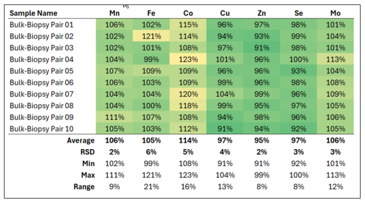
The results for each mineral summarized in (Table 1) show how well paired freeze-dried bulk-Tru-Cut biopsies compare. Perfect agreement would show a result of 100%. For all minerals, the range is from 91% to 123%, which is satisfactory since the smaller sample size would be statistically less likely to represent the bulk tissue. This much deviation is not abnormal in trace mineral analysis and should not greatly impact the clinical significance of the results.
Our normal 1 g samples and Tru-Cut biopsies showed good agreement across the mineral panel. Data is currently limited, but collection is ongoing to expand the number of observations for further statistical analysis. Preliminary results suggest that liver biopsies are suitable for trace mineral assessment.
To submit biopsy samples for analysis, collect 2 to 3 16-gauge Tru-Cut biopsies - see our previous LabNote (https://www.uoguelph.ca/ahl/ahl-labnote-19-liver-biopsy-procedure-cattle [1] ) on biopsy collection for more information. Place them into a plastic container or tube without gauze. Do not add any liquids (e.g. saline or fixative), as they may leach minerals from the tissue. Refrigerate or freeze and submit to the AHL. Results are reported on a dry weight basis with adjusted reference intervals where available.
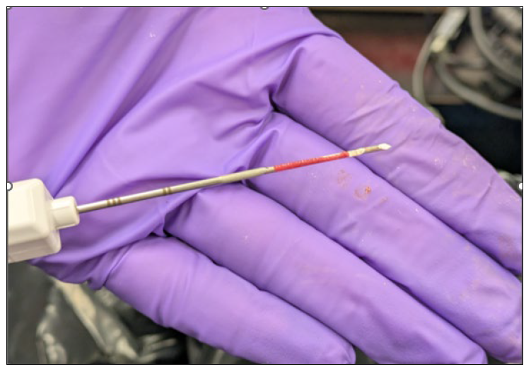
Figure 1. A freshly collected 16-gauge Tru-Cut biopsy sample showing a small core of liver tissue
Table 1. A summary of the relationship between 1 g “bulk” liver samples and Tru-Cut liver biopsies for the elements in the trace mineral panel. RSD = relative standard deviation = standard deviation ÷ mean average.
References
1. Herdt, T.H. and Hoff, B. The Use of Blood Analysis to Evaluate Trace Mineral Status in Ruminant Livestock. Vet Clin Food Anim 2011;27:255–283. https://www.sciencedirect.com/science/article/pii/S0749072011000065?via%3Dihub [2]
2. Radke, S.L., et al. Inductively coupled plasma mass spectrometry determination of hepatic copper, manganese, selenium, and zinc concentrations in relation to sample amount and storage duration. J Vet Diagn Invest 2020;32:103.. https://journals.sagepub.com/doi/10.1177/1040638719894988 [3].