Rebecca Egan, Anna Marom, Xi Xue, Davor Ojkic
Animal Health Laboratory, University of Guelph, Guelph, ON
AHL Newsletter 2026;30(3):14.
In the Ontario swine herd, porcine sapovirus (PoSaV)-associated diarrhea is frequently identified in young piglets, and most often with concurrent rotavirus infection. When histopathology is performed, atrophic enteritis typical of a viral cause is identified in many of these cases (Fig. 1). In order to better understand the impact of these pathogens, particularly in the face of co-infections, immunohistochemistry for rotavirus and in situ hybridization (ISH) for PoSaV can be used to assess for viral antigen in association with the intestinal lesion, with detection supporting a causal role in lesion development.
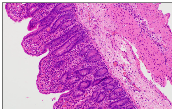
Figure 1. Small intestine from a newly weaned pig presenting with acute watery diarrhea, and atrophic enteritis demonstrated histologically by marked villous atrophy with blunting and fusion. PCR testing of feces was positive for rotavirus A (Ct 11.2), rotavirus B (Ct 27.6), rotavirus C (Ct 28) and porcine sapovirus (Ct 20.4), and negative for porcine coronaviruses. H&E stain.
In-house PCR testing for PoSaV has been available at the AHL since September 2023. Testing results were reviewed for 1,604 diagnostic samples representing 423 distinct cases received between September 6, 2023, and July 20, 2026, and cases with testing for porcine sapovirus and rotaviruses A, B, and C (RVA, RVB, and RVC) were selected. A case was classified as positive if at least one sample from the case was PCR-positive*. Overall, 395 (93.4%) cases were positive for at least one virus and only 28 (6.6%) were negative for all four viruses tested. Of the 233 PoSaV-positive cases, 209 (89.7%) had concurrent detection of at least one rotavirus, whereas only 24 (10.3%) were positive for PoSaV alone (Fig. 2). Among 190 PoSaV-negative cases, the most common infection patterns were RVA only, RVC only, and RVA–RVC co-infection (Fig. 4).
Investigation of the role of PoSaV in enteric disease in pigs is ongoing, but current information indicates that PoSaV does contribute to diarrhea, most often in neonatal pigs and occasionally in older nursing or weaning-aged pigs. Currently, the Ontario Animal Health Network has an ongoing project investigating the role of PoSaV in neonatal diarrhea complex, aiming to provide a better understanding of how PoSaV contributes to neonatal diarrhea cases in the Ontario herd. In situ hybridization (ISH) is being pursued for all PCR positive cases, which should provide insight regarding involvement of this pathogen in lesion development.
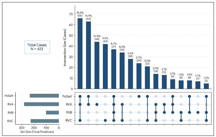
Figure 2. Co-infection patterns of PoSaV and rotaviruses A, B, and C among 423 positive cases tested at AHL since September 2023.
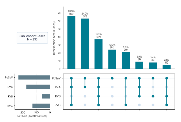
Figure 3. Rotavirus (RVA, RVB, RVC) co-infection profiles within porcine sapovirus (PoSaV)–positive cases (N = 233).
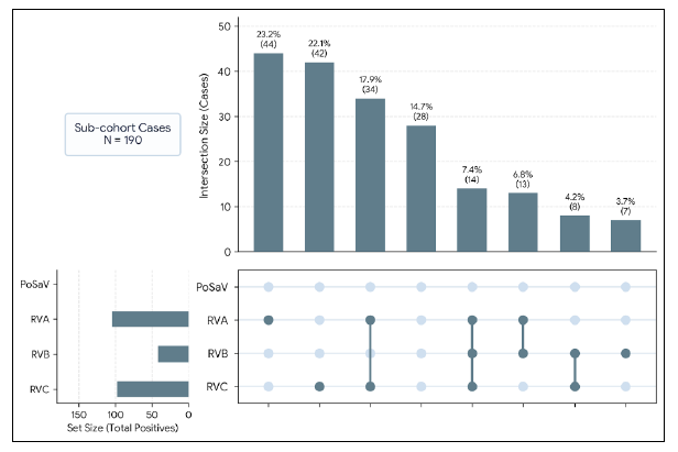
Figure 4. Rotavirus (RVA, RVB, RVC) infection profiles within porcine sapovirus (PoSaV)–negative cases (N = 190).
Sapoviruses are highly diverse viruses, with 21 genogroups currently identified in humans and animals. Eight genogroups have been reported in swine: GIII, GV, GVI, GVII, GVIII, GIX, GX, and GXI. PoSaV genotyping is based on sequence analysis of the major capsid protein gene, VP1. The first PoSaV isolate, the Cowden strain, was identified in 1980 and is now considered the prototype strain of genogroup GIII. This genogroup is the predominant and most widely distributed PoSaV lineage in swine worldwide, and all PoSaV detections in Ontario swine characterized to date have belonged to GIII. However, GIII exhibits substantial intra-genogroup diversity, encompassing multiple genotypes and continually giving rise to new genetic clusters as field strains evolve.
VP1 genotyping is now available at the AHL at a cost of $145 per sample (test code: psapose).
References
1. DeLay J, Ojkic D. Porcine sapovirus: An emerging pathogen contributing to swine diarrhea. AHL Newsletter 2023;27(1):15.
2. In: Diseases of Swine, 12th ed. Zimmerman JJ, Karriker LA, Ramirez A, Schwartz KJ, Stevenson GW, Zhang J, eds. Wiley-Black Sons; 2026. p 522-524.
3. Mizuno S, et al. Fecal metatranscriptomics and virus isolation reveal picornavirus diversity and evolution in Japanese wild boars and pigs. Sci Rep. 2026 Apr 9;16(1):20898.