Lisa Gordon
Animal Health Laboratory, University of Guelph, Guelph, ON
AHL Newsletter 2026;30(3):21.
An adult mixed-breed, male, neutered dog was presented with increased intraocular pressure (43 mm Hg) in the left eye. At this time, the patient was noted to be Ehrlichia spp. positive and was treated with a 30-day course of doxycycline. Two weeks later, an expansile conjunctival growth was observed dorsomedially (Fig. 1). A transpalpebral enucleation was performed, and the tissue was submitted for histological examination.
Histologically, the superficial lateral and medial sclera and surrounding periocular fibrous tissue and skeletal muscle are expanded and separated by extensive coalescing aggregates of macrophages, with clustered and scattered neutrophils, lymphocytes, plasma cells, multi-nucleated giant cells, and eosinophils admixed with cellular debris, fibrin, and hemorrhage on a background of plump fibroblasts and variable amounts of mature collagen. Within the inflammation there are numerous cross, longitudinal, and tangential sections of nematodes which range from 200 um – 250 µm in diameter (Fig. 2). Many of the parasites are hypereosinophilic (necrotic). The parasites have a ~5-10 µm thick, smooth, eosinophilic cuticle with ring-like circumferential cuticular ridges (annulations) that are apparent in the tangential and longitudinal sections (Fig. 2B inset). The organisms have a pseudocoelom and there are remnants of coelomyarian musculature which is segmentally thinned. The intestine is often eccentrically placed and small, and the reproductive organs contain abundant basophilic granular material. Occasionally, aggregates of the basophilic granular material are free within the inflammatory exudate. The conjunctiva is often segmentally attenuated and expanded by edema and large numbers of lymphocytes, plasma cells, and fewer macrophages and neutrophils.
There are accompanying pre-iridal and posterior corneal fibrovascular membranes, retinal detachment and segmental degeneration, and corneal neovascularization. The optic disc and the optic nerve are unremarkable. Neoplasia is not captured in section.
The globe was diagnosed with multifocal to coalescing granulomatous scleritis and periorbital fasciitis, steatitis, and myositis with intralesional degenerate adult filarid nematodes consistent with Onchocerca spp.
Causes of ocular helminthic infections in dogs include Onchocerca lupi, Thelazia spp., Ancylostoma caninum, Dirofilaria immitis, Angiostrongylus vasorum, Toxocara canis and Trichinella spiralis. The key diagnostic feature that differentiates Onchocerca spp. from other filarid nematodes is the presence of evenly spaced ring-like circumferential annular cuticular ridges seen on tangential/longitudinal section but not on cross-section (as are present in this case). Adult female Onchocerca spp. are the only nematodes that exhibit this cuticular pattern.
This parasite is not endemic to Canada and is rarely diagnosed locally except in dogs imported from endemic areas including southwestern United States (e.g. Arizona, New Mexico) and Europe. There are reports of development of Onchocerca nodules in dogs up to seven years after they were re-homed to a non-endemic region from an endemic one. This patient was adopted by the current owners approximately five years ago, and originated from Texas, USA.
Classic features of glaucoma (e.g. cupping of the optic disk) were not captured in section but are not always present histologically. Glaucoma in this patient is secondary and could be the result of altered flow due to fibrovascular membranes within the anterior chamber, due to compression of the filtration angle by the granulomas, or both.
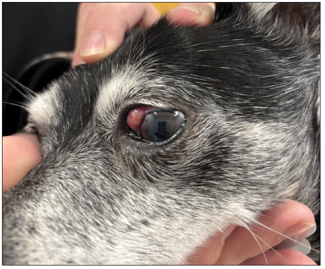
Figure 1. Conjunctiva, left globe. A smooth, pink-red, well-circumscribed, sessile mass arises from the dorsomedial conjunctiva of the left eye.
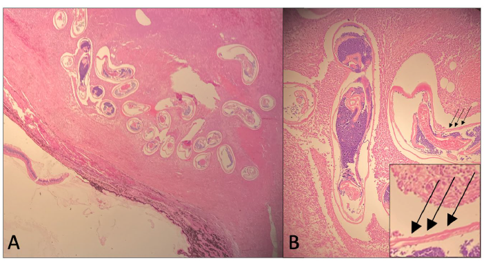
Figure 2. Sclera; H&E stain. 2A. 4X. Within the scleral inflammation there are numerous cross, longitudinal, and tangential sections of nematodes which range from 200 µm – 250 µm in diameter. Many of the parasites are hypereosinophilic. 2B. 10X. The parasites have a ~5-10 µm thick, smooth, eosinophilic cuticle with ring-like circumferential cuticular ridges (annulations) that are apparent in the tangential and longitudinal sections (inset, arrows). The organisms have a pseudocoelom and there are remnants of coelomyarian musculature which is segmentally thinned. The intestine is often eccentrically placed and small, and the reproductive organs contain abundant basophilic granular material.
References
1. McGarry JW, Carrozza R, Bradley C, Latrofa MS, Makepeace BL Otranto D. Onchocerca lupi in imported dogs in the UK: implications for animal and public health. BMC Vet Res. 2022 Feb 10;18(1):66.
2. Rojas A, Morales-Calvo F, Salant H, Otranto D, Baneth G. Zoonotic Ocular Onchocercosis by Onchocerca lupi. Yale J Biol Med. 2021 Jun 30;94(2):331-341.