CCMPS Researchers use Machine Learning to Transform Nanostructure Analysis
Drowning in Data at the Nanoscale
In biomedicine, soft nanoparticles are being explored as drug delivery vehicles to create more predictable, effective, and targeted medicinal therapies. For example, phytoglycogen is a naturally occurring complex sugar called a polysaccharide, that is structurally similar to glycogen, which is a molecule that animals use to store energy in the form of glucose.
Phytoglycogen acts like a nanoscopic sponge, holding a large amount of tightly bound water when fully hydrated. Due to its structure, it can also associate with several small bioactive molecules.
The polysaccharide has a tree-like structure, with a stiff inner core and a soft outer layer formed by the glucose branches of phytoglycogen, which can interact with the body. These properties, coupled with the biodegradability and non-toxicity of phytoglycogen, makes these particles desirable as nanocarriers.
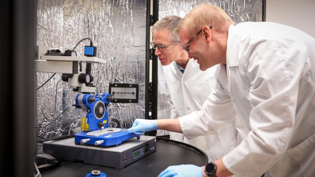
To better understand nanoscopic carriers, like phytoglycogen, material scientists and engineers use atomic force microscopy (AFM) force spectroscopy to measure a material’s stiffness (hard/soft), adhesion (stickiness), and deformation (squishiness). This force spectroscopy method determines these properties through the shape of force-distance curves, which are collected by measuring the force exerted on the tip of a tiny probe as it is pushed into and pulled away from a sample’s surface.
However, the resulting data analysis has historically been done manually. Researchers subjectively interpret thousands (or millions) of these force-distance curves, deciding on the location of nanoparticles and the interpretation of their physical properties. This method of analysis can be time consuming, introduce bias, and limit the reproducibility.
Using Machine Learning to Reliably Characterize Nanoparticles
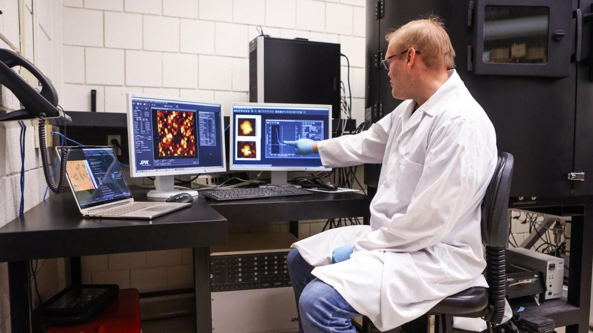
Postdoctoral scholar Dr. Benjamin Baylis, alongside Dr. John Dutcher in the Department of Physics, developed a machine learning-based framework to transform the way atomic force microscopy-force spectroscopy data is analyzed. An AFM force spectroscopy image can be thought of like a heat map for a material’s surface, revealing nanoscopic information on its structure and mechanical properties, where each pixel of an image corresponds to a single force-distance curve.
Dutcher and Baylis were interested in identifying the location of different materials and structures within a sample, and after a series of measurements, Baylis generated a set of physically meaningful features, such as stiffness and sample deformation, from each force-distance curve.
Ultimately, each machine learning model was trained on just two images, representing over half a million force-distance curves. Dutcher and Baylis assigned their machine learning models two tasks: to distinguish the soft phytoglycogen nanoparticles from the surface they were attached to, and to distinguish internal variations within the phytoglycogen nanoparticles.
The team also investigated the difference in accuracy between a 'supervised' machine learning model that was trained with labelled pixels, and an 'unsupervised' model that was trained with unlabelled pixels. For the supervised model, each pixel used in its training was pre-classified as corresponding to the soft outer layer of phytoglycogen, the stiff inner core, or the hard surrounding surface.
This labelling guides the machine learning model to learn how the meaningful features from the force-distance curves relate to the different regions in the sample.
Machine Learning Eliminates the Bottleneck in Analysis
Dutcher and Baylis found that after training on just those two images, both of their machine learning models were 99 per cent accurate in differentiating between the phytoglycogen nanoparticles and the hard surrounding surface.
The 'supervised' model was especially powerful in detecting the phytoglycogen nanoparticle's internal structure, successfully distinguishing stiffer inner regions from softer outer layers. The 'unsupervised' model could only distinguish obvious regions but struggled with subtle ones. They were then able to use the trained models to successfully distinguish between the different regions using new AFM measurements of phytoglycogen that the models had never seen before.
"We combined AFM force spectroscopy techniques and machine learning in a structured and physics-informed way to address a real bottleneck in data analysis, eliminating the need for manual analysis of overwhelming datasets,” says Dutcher.
"Even though our machine learning framework was based on a particular type of soft nanoparticle, phytoglycogen, it can be applied to other complex soft materials like polymers, gels, and even tissues, which has broader implications in materials engineering and biomedical applications."
The work of Dutcher and Baylis was highlighted on the front cover of an issue in the scientific journal Soft Matter.
This work was supported by a Natural Sciences and Engineering Research Council of Canada Discovery Grant
Baylis B. & Dutcher J.R. Machine learning approaches to quantify nanoscale variations in the mechanical properties of soft nanoparticles. Soft Matter. 2026. 22, 3143-3155. doi: 10.1039/D5SM00943J