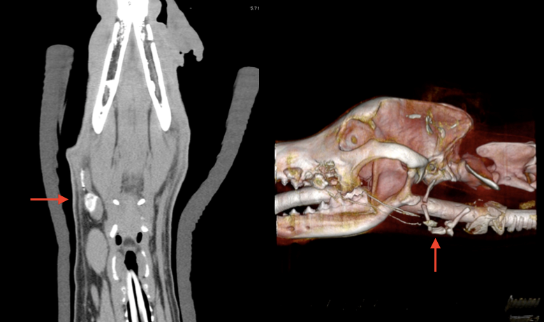
Over the past two years, I was able to collect data regarding Dr. Oblak’s canine oral tumour cases. These patients had contrast medium injected around an oral tumour, which allowed Dr. Oblak to visualize draining lymph nodes on CT. These lymph nodes are shown with red arrows on the CT and 3D reconstruction. Another contrast agent was then injected intraoperatively to identify and resect these lymph nodes. As a veterinary student, I’m very excited to be involved in such innovative clinical research and to be working with Dr. Oblak.