


AHL LabNote 33-Brain removal in field postmortems
Josepha DeLay, Maria Spinato, Andrew Brooks, Ken Bateman, Jeff Rau March 2015
Brain removal for histologic examination and microbiologic testing is valuable in animals that are euthanized or have died due to neurologic signs. Examination of brain is also important in cases of unexpected death. However brain removal is often avoided or overlooked during gross postmortem because of the physical challenge of this task, and important diagnoses may be missed. Similar issues arise if only 1-2 sections from brain are submitted, rather than the entire brain. An example is the difficulty in diagnosing listeriosis, which targets brainstem, if samples only from cerebral cortex are submitted for histopathology and microbiology.
If BSE, CWD, or scrapie is suspected, you must contact your local CFIA office and submit samples according to their instructions. If these diseases are included in the differential diagnosis but are not strongly suspected, consult your local CFIA District Veterinarian regarding your options before submitting the brain or head to the AHL for diagnosis. If rabies is suspected and there has been human involvement, please contact your local Public Health office. For rabies suspects with animal involvement only, please contact OMAFRA (1-877-424-1300).
Practitioners have 3 options for brain submission to the diagnostic lab:
1. Submit the intact head to the AHL. Brain removal and appropriate sampling will be performed at the lab. For head removal, disarticulate the atlanto-occipital joint beginning at the ventral aspect of joint. The joint is located at the level of the base of the ear and just caudal to the ramus of the mandible. Flexion and extension of the joint may aid in confirming its location. Cut through skin and soft tissue ventral and lateral to the joint with a large knife, sever the brainstem-spinal cord junction at the joint, and disarticulate by applying leverage between the articular surfaces of occipital condyles and the first cervical vertebra (C1).
The head may be delivered directly to the lab or shipped by usual courier methods. To prevent fluid leakage during transport, please ensure that the head is wrapped in 2 separate, individually sealed plastic bags and placed in a heavy cardboard box or plastic container. Several cold packs should be placed around the head to slow autolysis.
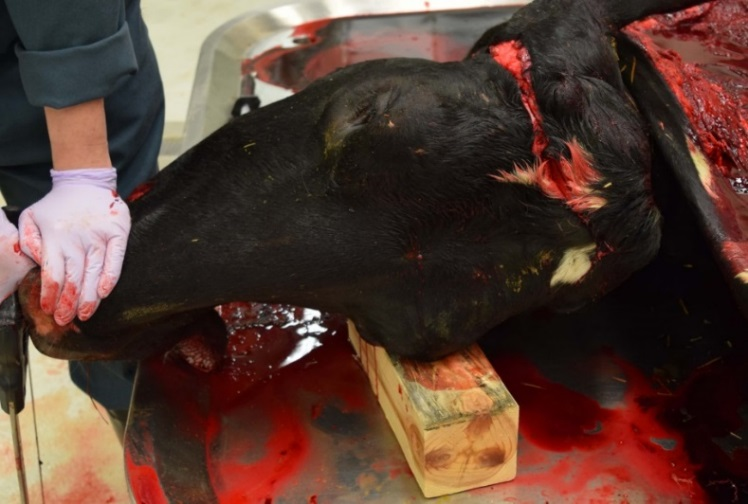
2. Brain removal using a lateral approach, cutting through the coronal plane. This method of brain removal can be easiest and safest for the operator, especially when removing brains from a large animal. With the animal in lateral recumbency and with the head still attached to the body, incise skin and soft tissue in a vertical line extending from just rostral to the base of the ear and caudal to the angle of the jaw (Figures 1, 2, and 3). Cut through the skull in this same plane using a rigid, flat cross-cut or other saw. Hacksaws are suitable for calves, foals, small ruminants, and large dogs, but the top band of a hacksaw may prevent deeper cuts required in more mature cattle, pigs, and horses. Separate the 2 segments of the sawn skull using a crowbar, or by levering the head over a block of wood (Figure 4). Rostral and caudal sections of brain can then be scooped out using a combination of manual dissection and transection of cranial nerves. Brainstem is severed at the obex to remove the caudal brain segment (Figures 5 and 6, and 7).
|
|
|
|
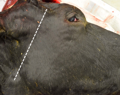
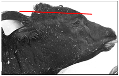
Figure 1. Landmarks (dotted line) for brain removal by lateral approach.
|
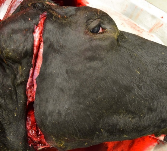
Figure 2. Skin and soft tissue incision for brain removal. Subsequent skull transection by saw cut will follow this same plane.
|
|
|
|
|
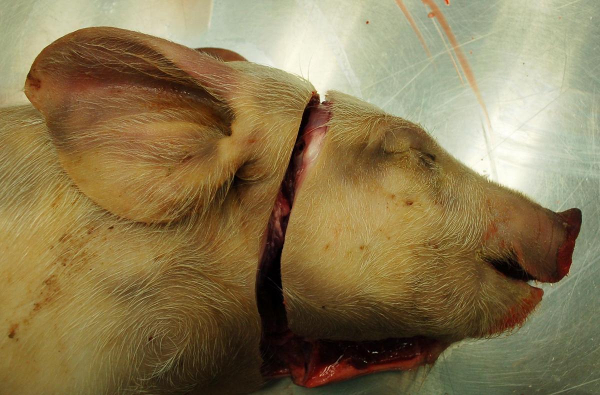
Figure 3.
|
Figure 4. Leverage application to separate sawn rostral and caudal skull segments. A crowbar or other lever may also be used.
|
|
|
|
|
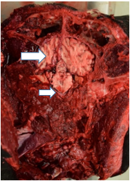
Figure 5. Rostral aspect of sawn skull, exposing cerebral cortex (large arrow) and pons (small arrow).
|
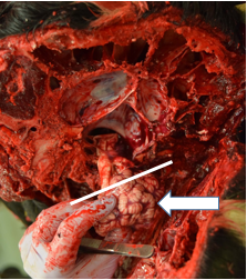
Figure 6. Caudal aspect of sawn skull, exposing cerebellum (arrow) and medulla, which is transected caudal to the obex (line) for removal. |
 |
|
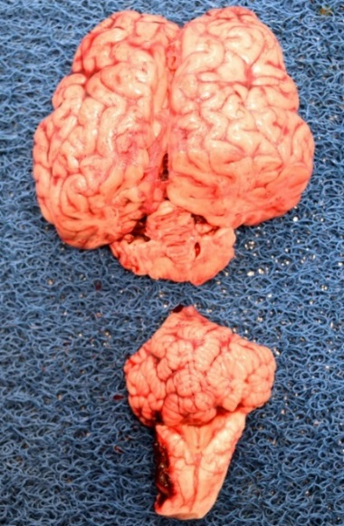
| Figure 7. Brain removal in rostral and caudal sections. These may be sagittally sectioned, with half of each section fixed in formalin for histopathology, and the remaining halves stored fresh or frozen for microbiologic tests. |
3. Brain removal using dorsal approach, removing the calvaria (skull cap). This technique uses an axe to open the skull and requires that the carcass is placed on a firm but shock-absorbing surface, such as dirt / pasture or straw pack (NOT concrete). BE SAFE – ensure that other people and animals are well away from the area, and the axe operator should be wearing appropriate footwear (steel-toed boots) and protective eyewear. Keep the head attached to the body for brain removal. Using the landmarks outlined (Figure 8) and an axe, remove the skull cap by directing the axe cut from the poll toward the front of the skull, just dorsal to the eye. In most cases, this will require several axe strikes. An alternative to consider is the use of a battery-powered reciprocating saw (“Sawzall”) with a 9 or 12 inch blade (depending on the age of the animal) with 5 or 6 teeth per inch. The dorsal aspect of cerebral hemispheres and cerebellum may be severed in the process, however sufficient brain should remain intact for sampling (Figure 9). With the skull cap removed, scoop the brain out of the skull, cutting cranial nerves at the ventral aspect the brain. Remember to include cerebellum and brainstem, which will be caudal to the exposed cerebral cortex and may still be obscured by a section of occipital bone.
As for the brain removed by the lateral approach described above, make a mid-sagittal cut in the brain once it is removed from the skull. Place one half in a large Whirl-Pak bag and chill. Make several transverse, partial-thickness incisions in the remaining half, and place in formalin – the incisions will allow better fixation of a large brain. Submit both fresh chilled and formalin-fixed brain to the diagnostic lab.
 |
 |
| Figure 8. Location of cut to remove skull cap. |
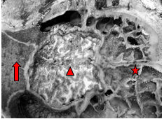
Figure 9. Dorsal aspect of skull following removal of skull cap. Cerebral cortex is visible (arrowhead); cerebellum and brainstem are ventral to an intact segment of occipital bone (arrow). Frontal sinus and ethmoid turbinates are visible to the right (star). |
File attachments
| Attachment | Size |
|---|---|
| 947.15 KB |