


AHL LabNote 54-Blackhead (histomoniasis) in small turkey flocks
Marina Brash, Lloyd Weber
Blackhead disease, also known as histomoniasis and enterohepatitis, is a disease that is not infrequently diagnosed at the AHL in late summer and fall in small turkey flocks and, because there are no approved preventive or treatment medications, mortalities can persist resulting in very high losses.
Blackhead is caused by a protozoan parasite, Histomonas meleagridis, which is carried by the common poultry cecal worm Heterakis gallinarum, found in the ceca of chickens and turkeys. This protozoan organism is fragile and cannot live outside the bird host for long but can survive for long periods of time in the environment when in the cecal worm or its eggs. Earthworms can also carry infected cecal worm eggs and are important in transmitting blackhead. Flies, beetles, including darkling beetles, grasshoppers, sowbugs, and crickets can also serve as mechanical vectors. Cecal worms and eggs can be also be transferred via contaminated manure on equipment and shoes.
Typically, outbreaks in turkeys begin with the ingestion of infected earthworms or cecal worms or eggs from contaminated soil or concrete-floored pens where flocks of chickens or turkeys have been raised. Wild grouse and quail may also carry the infection to turkey yards. In turkeys, bird-to-bird transmission, through direct cloacal contact or with infected droppings, can contribute to maintenance of the outbreak. Chickens, pheasant, partridge, and peafowl are also susceptible.
Once the H. meleagridis organisms are ingested, they travel to the cecum, penetrate the mucosa, multiply, resulting in necrosis, inflammation, and thickening of the cecal wall, enter the bloodstream and travel to the liver inciting necrosis and inflammation. In the intestine, interaction with coccidia and bacteria including Clostridium perfringens and E. coli increases the severity of disease with more tissue damage. Turkeys can show clinical signs of illness 7-12 d post-ingestion including anorexia, drowsiness, ruffled feathers, drooping of the wings, placement of the head down and close to the body or tucked under the wing, and the excretion of yellow feces described as sulfur-yellow droppings (Fig 1). The head may or may not be cyanotic. Death follows shortly thereafter.
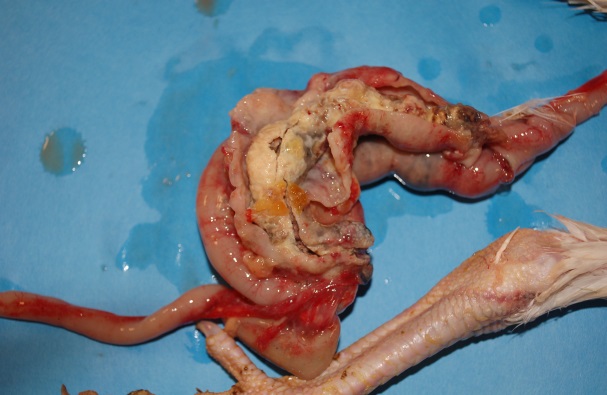
At postmortem, lesions are characteristic with enlargement of the cecal pouches and marked thickening of cecal walls with thick caseous cores (Fig .2). Extension of inflammation through the cecal serosa will result in localized peritonitis. The liver is enlarged, with multiple circular depressed areas of necrosis circumscribed by raised yellow rings described as targetoid lesions (Fig. 1). Lesions can also be found in other organs, including kidneys, bursa of Fabricius, spleen, lungs, pancreas, and proventriculus.
Histopathology can be conducted to confirm the diagnosis as the histomonad organisms will be present in cecal and liver tissues.
There are no approved preventative or treatment medications, therefore prevention of this disease is the key and the approach is multipronged:
- Chickens and turkeys should be raised in separate areas. Turkeys should not be reared where chickens have been housed previously. Free-roaming chickens and turkeys may visit areas frequented by wild turkeys, and gamebirds including pheasants, partridge, grouse, and quail and are much more likely to pick up cecal worms.
- If there is a history of blackhead on the farm, free-roaming domestic fowl must either be relocated to new fenced pasture areas or moved indoors to pens that have not previously housed chickens or turkeys, to prevent contact with intermediate hosts and contaminated soil or pen floors. Concrete-floored pens are preferred to dirt floors.
- Straw and bedding storage areas should be bird-proof to prevent fecal contamination. New litter should not be stored/contaminated with old litter. New litter should not be stored where poultry has been previously housed.
- Control mechanical vectors including earthworms, beetles, including darkling beetles, flies, and other arthropods and manure-contaminated equipment and shoes.
- Routine deworming to control cecal worm infestations will require a veterinary prescription as there are no medications approved for use in turkeys. The cecal worm burden can be evaluated by inspection of cecal contents during postmortem examination for these small nematodes. Ascarid and cecal worm eggs can be differentiated by size, so fecal flotations are another more feasible option.
- If the flock is experiencing higher than normal mortality, contacting your veterinarian as quickly as possible for a postmortem is essential as there has been some success with moving the clinically normal turkeys inside or to another pen away from the contaminated environment and dimming the lights to reduce litter pecking. Moving sick turkeys is not recommended as they will only contaminate the new site.
- After the turkeys are removed from the barn, the contaminated litter must be removed. If the barn has concrete or wood floors, washing and disinfecting the barn works well. Hydrated lime, available at farm store outlets, offers an alternative to disinfection for barns with dirt floors. Spread 40 pounds of hydrated lime over 1,000 square feet of dirt floor and cover with straw or shavings. Please refer to the manufacturer’s handling instructions as hydrated lime is both corrosive and toxic.
- If excessive mortality in mature turkeys is being experienced and it is possible to move up the processing date, then that should be considered for the remaining healthy birds. However, it is important to advise the producer that if at processing, the turkeys are found to be dehydrated and/or in poor body condition and/or have grossly affected internal organs, they will be condemned.
Figure 1: Turkey: Sulfur-yellow droppings on the underside of the tail feathers, typical targetoid liver lesions
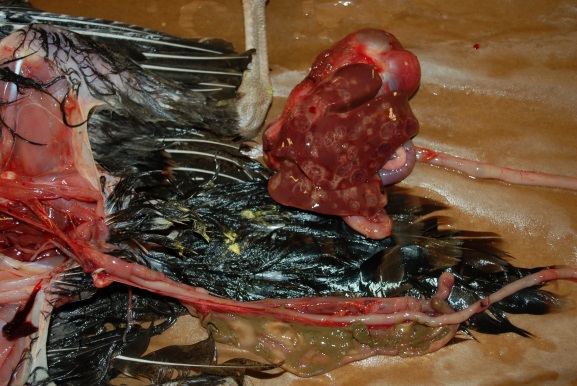
Figure 2: Turkey: Typical cecal lesions
File attachments
| Attachment | Size |
|---|---|
| 419.42 KB |