Bovine Diarrhea Diagnostic Plan - July 2018
A. History:
· Provide information on herd size, animal age (very important! Re set up of bacteriology testing) , numbers of animals affected, onset/duration of problem, vaccination history, and any treatment administered.
B. Selecting and submitting samples:
· For best results, select acutely affected, untreated animals for sampling, OR submit feces from same.
· Live acutely affected young ruminants (<40 kg) may be submitted for postmortem examination. **Please call the lab first to arrange, and ensure arrival PRIOR to 15:00 h.
Live animals must be transported humanely.
· Rapid tissue fixation of gut sections for histology is critical, preferably <10 minutes after death.
· Since intestinal lesions may be segmental, always collect multiple intestinal tissue samples for histology,
preferably 3 segments each of ileum and jejunum, and one each of duodenum, cecum, colon and stomach.
Partially snip open one end of 1-2 cm gut sections with scissors to expose mucosa prior to immersion in formalin.
· Please request specific tests, and submit separate tissue samples, in separate labeled Whirl-Pak bags, for each lab section and test requested. Alternatively, ask for pathologist to select tests as appropriate. (sampling fee)
C. Samples required:
Bovine enteritis:
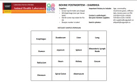
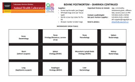
Histology: Formalin-fixed sections of esophagus, forestomachs, abomasum, mesenteric lymph node, duodenum, jejunum, ileum with Peyer’s patch, spiral colon and cecum, including any area with gross lesions. Also include routine tissues, e.g., filtering organs liver, kidney, spleen, lung. Remember to collect multiple sections from jejunum and ileum.
• Immunohistochemistry is available on formalin-fixed tissues for BVDV, BCV, bovine rotavirus.
(Note that BVDV IHC is NOT available for routine PI screening – PCR is the test of choice – pooling is available).
Bacteriology: Feces, small and large intestine, mesenteric lymph node, any other tissues with visible lesions – routine culture.
• Mycobacterium paratuberculosis (Johne’s disease): PCR (intestine, mesenteric lymph nodes, feces).
Virology:
• BVDV: PCR (EDTA blood for acute BVD, serum for PI; tissues including ileum with Peyer’s patch, spleen, mesenteric lymph node, esophagus); For further details of BVDV testing, see LabNote 1 - Summary of BVDV testing at the AHL
• Rotavirus A and B; Coronavirus: PCR (feces, ileum/colon).
Serology: Paired sera for BCV. For acute BVD, submit paired sera for both type 1 and type 2 BVDV VN tests.
Parasitology: Feces for Cryptosporidium sp.(sucrose wet mount – semi quantitative), coccidia, gastrointestinal nematodes (GIN) (fecal flotation).
Please note: Bovine neonatal enteric panel – includes bovine coronavirus, rotavirus ruminant group A and B – PCR (rocopcr), sucrose wet mount (sucwt), bacterial culture, food/fiber producing animals (other than swine) (cultf, bsetup). Feces needs to be split into 3 separate sterile vials for distribution to the 3 lab sections involved. If done at AHL, an extra splitting charge will be required.
See Bovine Postmortem sampling guide - Diarrhea