


Feline pseudomycetoma; an uncommon presentation of Microsporum canis infection in cats
Emily Rätsep
Animal Health Laboratory, University of Guelph, Guelph, ON.
AHL Newsletter 2025;29(4):30.
A female spayed, 12-year-old Persian cat had a history of slowly expanding round dermal masses that had been present and visible to the owner for a period of 1-2 years. The rate of growth was slow, though ulceration of the skin overlying the two masses was observed, leading to their removal. Both tissues (haired skin samples) were submitted as biopsies at the Animal Health Laboratory. On tissue sectioning, the nodules contained yellow granular purulent material.
Histologic examination revealed ~1.1 cm x 2.0 cm to 1.2 cm x 3.3 cm ulcerated masses expanding and effacing the dermis. These were comprised of sheets of epithelioid macrophages and multinucleated giant cells, admixed with viable and degenerate neutrophils (Fig. 1). These sheets of inflammatory cells were centred around radiating Splendore-Hoeppli material, and oval to polyhedral granular structures that in some areas appeared to form ovoid to elongated fungal spores or hyphal masses (Fig. 2). Both masses were confirmed to be inflammatory reactions to fungal elements as opposed to neoplasia. These histological features are consistent with a diagnosis of dermatophytic pseudomycetoma.
Dermatophytic pseudomycetomas with subsequent pyogranulomatous reaction are a rarely-reported invasive fungal infection, that have been observed in humans and animals - felines in particular. In cats, this is typically a rare presentation of a Microsoporum canis infection. While feline dermatophytosis due to infection with M. canis is common, mycetoma formation is not. Typical M. canis infections are non-invasive, with restriction of the fungal elements to colonized surface skin. When involvement of the deeper tissues occurs, an inflammatory mass can result. Interestingly, mycetoma formation is almost exclusively reported in Persian cats. While the underlying cause of this reported increased incidence is uncertain, an inherited susceptibility is suspected.
Dermatophytic pseudomycetomas typically form nodular, non-painful, non-pruritic firm masses, usually located on the trunk, flanks or tail; concurrent superficial dermatophytosis is common. These masses can ulcerate and occasionally, infections can form a fistula with exudation of the purulent and fungal material to the exterior surface of the skin. Occasionally, the fungal elements in these dermal masses can invade the deeper subcutis, complicating both treatment and possible surgical resection.
As M. canis is a zoonotic pathogen, awareness of the possible different presentations possible for infection with this fungal species is useful, especially in clinical practice.
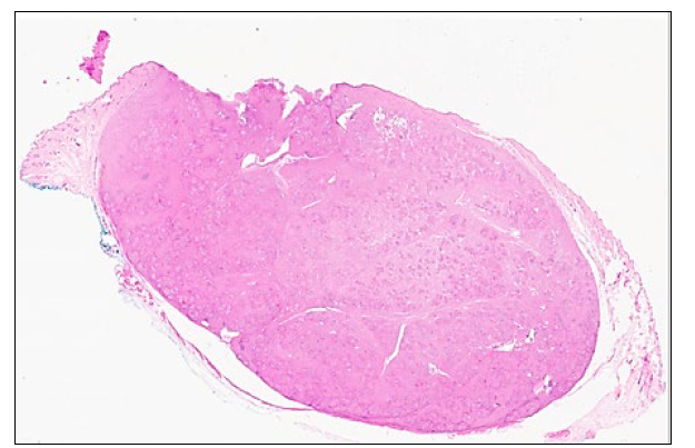
Figure 1. Skin from a 12-year-old cat; there is a focal mass of inflammatory cells elevating the partially ulcerated skin surface (2x). H&E stain.
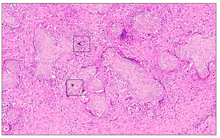
Figure 2. Skin from a 12-year-old cat. Multinucleated giant cells, macrophages and degenerate neutrophils are centered on Splendore Hoeppli material (<) surrounding fungal elements (*). 20x.
H&E stain.
References
1. Barrs VR, et al. Invasive fungal infections and oomycoses in cats. 1. Diagnostic approach. J. Feline Med. Surg. 2024. 26:1-22.
2. Hnilica KA. Fungal skin diseases. In: Small Animal Dermatology, 4th ed. Hnilica KA, Patterson AP, eds. Saunders Elsevier, St. Louis, MO, 2011: 102.
3. Kano R, et al. Confirmed case of feline mycetoma due to Microsporum canis. Mycoses 2008;52:80-83.