


Gangrenous dermatitis in commercial poultry
Emily Martin and Emily Brouwer
Animal Health Laboratory, University of Guelph, Guelph, ON.
AHL Newsletter 2024;28(1):28.
In the fall of 2023, the AHL received multiple cases of broiler breeder chickens with increased mortality and skin lesions. The skin changes varied from mild congestion and subcutaneous edema to marked red-purple discolouration of the skin, edema, and crepitus (Fig. 1). Bacterial culture of subcutaneous swabs identified various combinations of C. septicum, C. perfringens, Staphylococcus aureus, E. coli and E. cecorum. The presence of Clostridium sp. was consistent with a diagnosis of gangrenous dermatitis.
Gangrenous dermatitis can occur in both chickens (4-5 weeks of age) and turkeys (13-16 weeks of age) that are approaching market age. The economic impact occurs due to increased mortality, increased condemnations, and carcass downgrading. Mortality is usually 1-5% higher in affected flocks compared to unaffected flocks, but can occasionally exceed this level (i.e., up to 60%).
Flocks present with a sudden increase in mortality. Clinically, the birds are depressed, off feed, weak, ataxic and laterally recumbent. The most significant locations for skin lesions include the breast, back, abdomen, thighs, wings, and tail (turkeys). The skin lacks feathers can be dry or moist and is discoloured dark red or green. Affected skin can have a range of lesions including edema, emphysema, and multifocal to coalescing hemorrhages that can extend into the underlying musculature. Carcasses rapidly autolyze. Microscopically, necrosis primarily occurs in the epidermis and dermis but can extend into the subcutaneous tissues and underlying skeletal muscle (Fig. 2). The subcutaneous tissues have accumulation of serofibrinous exudate and emphysema. There are Gram-positive rods noted throughout all skin layers and muscle with minimal inflammatory response.
The primary bacteria involved are either C. septicum, C. perfringens type A, or both. These bacteria produce toxins resulting in the tissue necrosis described. Numerous other aerobic or anaerobic bacteria can be isolated in combination with these clostridial species.
Environmental conditions and immunosuppression can interact to predispose to development of this disease. Environmental conditions include skin lesions (i.e., trauma due to cannibalism or fighting), stocking density, wet litter, poor ventilation, water leaks, and contamination (i.e., feed, water, equipment, vaccines). Immunosuppressive agents include IBDV, CAV, reovirus, IBHV, and HEV (turkeys).
The pathogenesis is not well understood and there are two theories for the introduction of bacteria. The first theory is skin trauma with external seeding of bacteria. The second is intestinal overgrowth of bacteria, loss of intestinal integrity, and systemic distribution of bacteria. Potential rule outs include contact dermatitis, mycotic dermatitis, bacterial cellulitis (non-clostridial), scabby hip (broilers) and focal ulcerative dermatitis (turkeys).
A presumptive diagnosis can be based on clinical signs, postmortem lesions, and histopathology. Subcutaneous swabs for aerobic and anerobic culture can be used to confirm this diagnosis. AHL
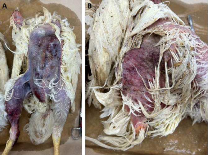
Figure 1. Skin lesions suggestive of gangrenous dermatitis (A: Breast. B: Lateral thigh)
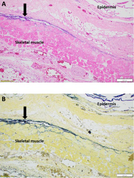
Figure 2. Histologic lesions of gangrenous dermatitis.
A: H&E stain. Necrosis and edema of dermis, subcutis and muscle including a narrow purple line of bacterial growth (arrow).
B: Gram stain showing blue staining of Gram-positive rod bacteria consistent with clostridial species (arrow).
References
1.Gornatti-Churria CD, et al. Gangrenous dermatitis in chickens and turkeys. J Vet Diagn Invest 2018;30:188-196.
2.Li G, et at. An outbreak of gangrenous dermatitis in commercial broiler breeders. Avian Pathology 2010;39:247-253.
3. Lighty ME, et al. Incidence of clostridial dermatitis (cellulitis) and factors for development of the disease in turkeys. L Appl Poult Res 2016;25:104-112.
4. Opengart K. Gangrenous Dermatitis. In: Swayne DE, ed. Diseases of Poultry, 14th ed., Vol II. Wiley Blackwell, 2020:980-985.