


Necrohemorrhagic tracheitis in swine: diagnostic investigation
Funding for this project is subsidized by the Swine Health Information Center (SHIC) and covers fees for specific testing on tracheal samples. Clients are responsible for test costs for routine respiratory pathogens.
To qualify for subsidized testing, the case definition must be met (below) and appropriate samples must be submitted to the AHL.
Please contact the AHL if you would like to submit a case through this project, or if you have any questions: Dr. Josepha DeLay 519-824-4120 ext. 54576 jdelay@uoguelph.ca
Case definition: Hogs (including breeding stock), with age range 14-30 weeks and weight range 150-350 lbs (70-160 kg), with acute onset of ‘honking’ cough in a low % of animals, progressing to dyspnea and mortality/euthanasia. Necropsied animals feature edema and hemorrhage within tracheal submucosa, resulting in marked luminal impingement.
Desired samples and other information :
Clinical history, including % morbidity / mortality, approximate weight of hogs, herd genetics
Gross images of cervical region and pluck at time of necropsy
Samples from 1- 3 affected pigs:
Intact plucks (trachea from larynx to bifurcation, and both lungs) (minimum sample required)
Tracheas should be partially opened during the field necropsy to confirm lesions compatible with necrohemorrhagic tracheitis (Figure 1). Further dissection and sampling of trachea and lung for diagnostic testing and lesion documentation will be done at the diagnostic laboratory.
Bilateral caudal deep cervical and costo-axillary lymph nodes
Lymph nodes should be submitted intact and fresh, and labelled (right and left, anatomic site).
Control animals: Submission is encouraged of plucks and lymph nodes as above from 2-3 unaffected herdmates dying or euthanized during the same timeframe, potentially due to other causes, and without gross lesions of necrohemorrhagic tracheitis.
Fresh and formalin-fixed spleen, stomach, ileum, and heart.
Oral fluids from all affected pens, 2 unaffected pens in affected barn, and 2 pens in unaffected barns.
Serum from 5 animals in each of 1 representative affected pen, 1 unaffected pen, and 1 pen in an unaffected barn.
Environmental control records for affected barns for 2 weeks prior to outbreak, targeting sudden fluctuations in temperature or humidity; comment regarding barn air quality.
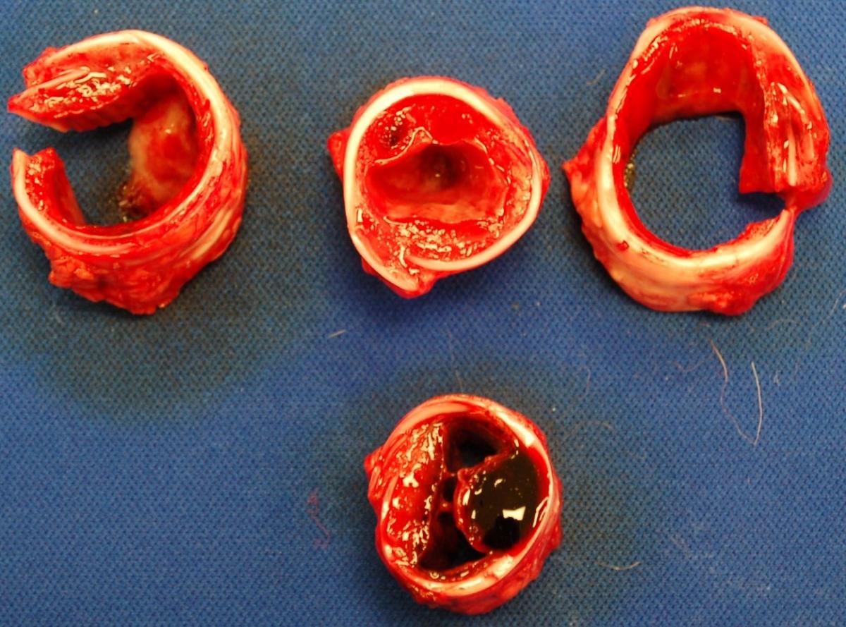
Figure 1. Tracheal cross-sections from multiple finisher pigs with necrohemorrhagic tracheitis. Note severe submucosal hemorrhage and edema, with resulting reduction in tracheal luminal area.
Client costs: gross exam, histopathology, bacterial culture setup and 1-3 samples (lung), Influenza A virus PCR (lung x3), PRRSV / PCV / M.hyopneumoniae PCR (pooled samples - discounted)
SHIC subsidy: bacterial culture for 3 samples (superficial and deep tracheal mucosa, spleen), Influenza virus PCR (tracheal mucosa x3), Influenza IHC (if required), 15% subsidy if PRRSV / PCV / M.hyopneumoniae PCR done based on gross / histologic lesions.
File attachments
| Attachment | Size |
|---|---|
| 718.28 KB |