


Necrotizing pancreatitis in guinea fowl associated with adenovirus infection
Andrew Brooks, Emily Martin, Alex Weisz
Animal Health Laboratory, University of Guelph, Guelph, ON; Smith and Weisz Poultry Veterinary Services, Kitchener, ON (Weisz).
AHL Newsletter 2024;28(1):27.
In January 2024, the AHL received a submission from a flock of guinea fowl experiencing elevated mortality. The mortality was affecting younger birds approximately 1-2 weeks of age. The on-farm postmortem examinations revealed gross lesions of necrosis and inflammation in the pancreatic tissues of the affected birds.
Histologically, all sections of pancreas exhibited severe, multifocal to generalized necrosis, with interstitial inflammation (Fig. 1A). There was also atrophy and loss of the pancreatic parenchyma. In some sections of pancreas, the necrosis was confluent. There were exudates of fibrin on the pancreatic surface and in some sections, there was generalized inflammation with infiltrates of lymphocytes and plasma cells expanding the interstitium. Throughout all of the pancreatic lesions, there were numerous large amphophilic intranuclear inclusion bodies consistent with adenovirus (Fig. 1B).
In guinea fowl, fowl aviadenovirus infections have been associated with erosions in the gizzard, ventriculitis and pancreatitis, and outbreaks of necrotizing pancreatitis have been reported in young guinea poults. A recent investigation of outbreaks of mortality and pancreatitis in guinea fowl flocks in Europe identified fowl adenovirus type 1 (FAdV-1) as the etiologic agent. In that investigation, clinical disease and mortality were highest in younger birds between 1-3 weeks of age, and mortality reached as high as 10-25%. The diagnosis of this disease in guinea fowl is based on the gross and histological lesions, including characteristic inclusions in the pancreas, as well as detection of FAdV-1 by PCR. AHL
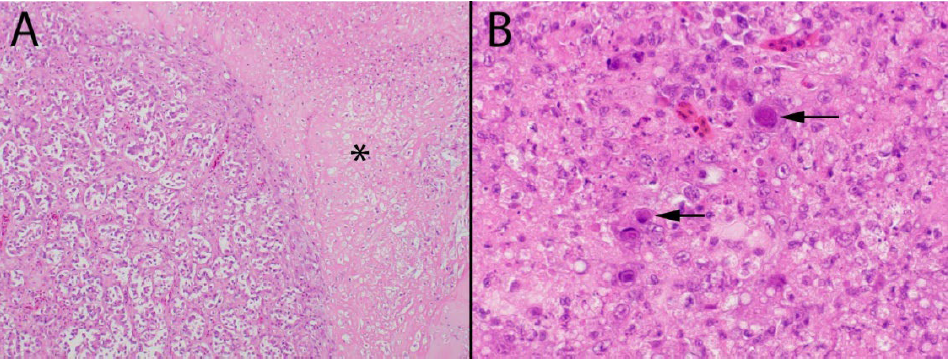
Figure 1. Lesions in the pancreas of guinea fowl associated with adenovirus infection. (A) There is extensive pancreatic necrosis (*) with inflammation, atrophy and loss of parenchyma. (B) There are numerous intranuclear inclusion bodies consistent with adenovirus (arrows). H&E stain.
Reference
1. Croville G, et al. Detection and typing of a Fowl Adenovirus Type 1 agent of pancreatitis in guinea fowl. Avian Dis 2021; 65(3):429-437.