


Update on porcine sapovirus detection in Ontario swine
Davor Ojkic, Josepha DeLay
Animal Health Laboratory, University of Guelph, Guelph, ON.
AHL Newsletter 2024;28(1):25.
In-house PCR testing for porcine sapovirus (PSaV) has been available at the AHL since September 2023. Results to date from Ontario herds are similar to those described in other geographic areas in North America, with PSaV-associated diarrhea most frequently identified in nursing piglets around 10 days of age, often associated with rotavirus co-infection.
Between September 2023 and February 2024, a total of 83 samples from 32 Ontario herds were tested by PCR for PSaV. Of these, PaSV nucleic was detected in 30 samples from 16 herds. The age range of PSaV-positive pigs was 3-28 days, with an average age of 9 days. Histopathology was carried out for 13 of the 16 PSaV-positive herds, and atrophic enteritis typical of viral enteritis was identified in 12 of the 13 herds (Fig. 1). In situ hybridization (ISH) for PSaV in 2 herds confirmed viral presence within intestinal lesions (Fig. 2). Rotavirus PCR was carried out on each of the PaSV PCR-positive samples, and 17/30 (57%) samples had concurrent infection with rotavirus A, B, and / or C. PCR for porcine enteric coronaviruses (PEDV, TGEV, PDCoV) was carried out on 9 of the PSV-positive samples that were also tested for porcine rotavirus, and coronaviruses were not detected in any of the samples.
These findings reinforce the importance of testing for multiple viral pathogens in nursing and some older pigs with diarrhea. Although the full significance of PSaV to the Ontario swine herd remains to be determined, current information indicates that the virus does contribute to diarrhea predominately in neonatal pigs, but also occasionally in older nursing or weaning-aged pigs. AHL
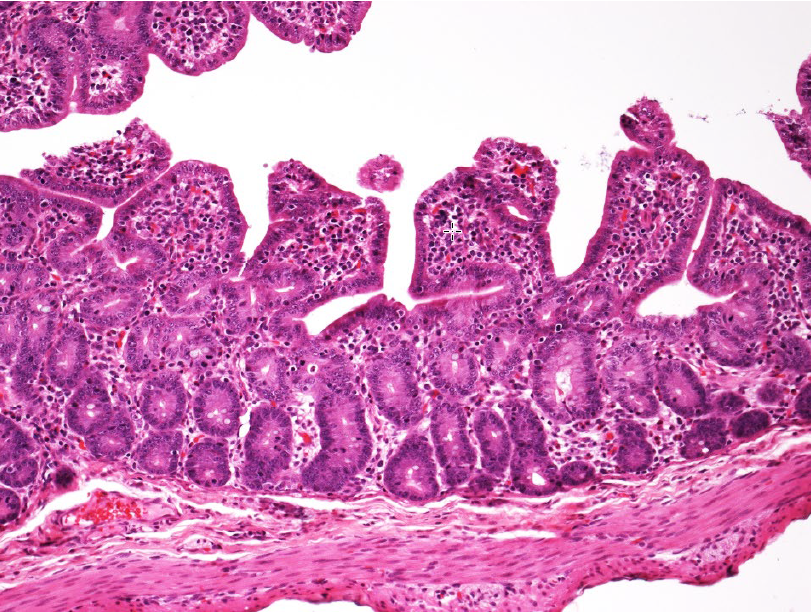
Figure 1. Atrophic enteritis in a 21-day old pig. Villi are short and blunted, and cuboidal to jumbled epithelium covers villus tips. PSaV nucleic acid was detected in feces by PCR (Ct 16.82). H&E stain.
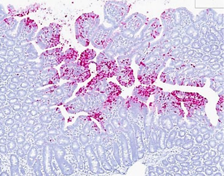
Figure 2. PSaV RNAscope (in situ hybridization), small intestine of same pig as in Figure 1. PSaV nuclei acid (pink chromogen) is detected in small intestinal mucosa, predominately in epithelial cells at villus tips and in lamina propria. Although porcine rotavirus nucleic acid was also detected by PCR in feces from this group, rotavirus antigen was not detected in association with histologic lesions of atrophic enteritis. Taken together, these results support PSaV as the primary etiology for atrophic enteritis and diarrhea in this group. Photo courtesy of Dr. R. Derscheid, Iowa State University.
References
1. Nagai M, et al. Porcine sapoviruses: Pathogenesis, epidemiology, genetic diversity, and diagnosis. Virus Res 2020; https://doi.org/10.1016/j.viruses.2020.198025
2. Shen H, et al. Genetic characterization of porcine sapoviruses identified from pigs during a diarrhea outbreak in Iowa, 2019. Transbound Emerg Dis 2022;69:1246-1255.
3. Wang L, et al. Genetically divergent porcine sapovirus identified in pigs, United States. Transbound Emerg Dis 2020;618-28.