


COMPANION ANIMALS
A case of feline paraneoplastic alopecia
Jan Shapiro, Andrew Brooks
An 11-year-old domestic shorthair cat was submitted for postmortem examination to the Animal Health Laboratory in Kemptville. The owner had presented the cat to a veterinary clinic with the complaint that it had been ill for a couple of weeks and had hair loss. Given the very poor condition of the cat, the veterinarian recommended euthanasia.
Postmortem examination of the cat revealed dramatic alopecia, affecting at least 30% of the body (Fig. 1). The affected areas included the base of the ears, the intermandibular area, posterior and ventrolateral aspect of the mandibles and neck, pectoral area, axillae, ventrolateral thorax and abdomen, inguinal areas, anus and perineum, the underside of the tail, and large patchy areas of all 4 legs including the digits. The alopecic skin was smooth, red and often shiny, and the ventral abdominal skin was wrinkled. The hair adjacent to the alopecic areas was easily epilated.
On internal examination, the liver had numerous discrete tan-pink oval-to-stellate masses of 0.1 x 0.1 cm to 0.8 x 1.2 cm. The masses bulged from the capsular surface of the liver, and were present in the parenchyma of all lobes. The left thyroid gland was 4-fold heavier than the right, and normal thyroid tissue was compressed and displaced to one pole by an encapsulated tumor mass. The heart had moderately severe biventricular dilation, and the lungs were mottled pink and red, with mild rib impressions.
Histologic examination of skin sampled from multiple sites showed severe follicular atrophy, mild parakeratosis, mild multifocal acanthosis, and localized bacterial pyoderma. The liver tumor was diagnosed as a low-grade cholangiocarcinoma, with multinodular metastasis to the lungs. Additional diagnoses included thyroid adenoma and multifocal myocardial fibrosis.
The histopathology of the skin was consistent with that reported for feline paraneoplastic alopecia, in this case associated with malignant neoplasia of the bile ducts.
Cutaneous paraneoplastic syndromes are dermatoses associated with internal neoplasms, usually malignant. Feline paraneoplastic alopecia (FPA) is a rare dermatosis of aged cats that is characterized by symmetrical ventral body and limb alopecia, and has been associated with pancreatic or bile duct carcinoma. The pathogenesis is not known, but the alopecia is usually reported as acute in onset and progressive, occurring over as short a period as 2 wk or as long as a few months. Hair is lost from the ears, ventral chin, neck, thorax, abdomen, and legs, and the remaining hair epilates easily. As in the case reported here, areas of alopecia are often smooth, red and shiny, and pruritus is not typically reported. Alopecia may precede, be concurrent with, or follow the diagnosis of the tumor. AHL
References
Gross TL, et al. Atrophic Diseases of the Adnexa. In: Gross TL, et al. Skin Diseases of the Dog and Cat, 2nd ed. Oxford: Blackwell, 2005.
Mauldin EA, Peters-Kennedy J. Integumentary System. In: Maxie MG, ed. Jubb, Kennedy, and Palmer’s Pathology of Domestic Animals, 6th ed. St. Louis, MO: Elsevier, 2016.
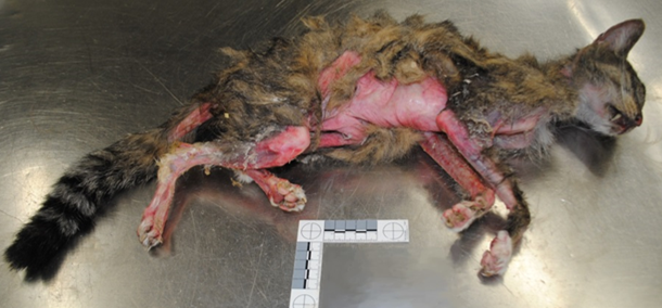
Figure 1. 11-year-old domestic shorthair cat with feline paraneoplastic alopecia associated with cholangiocarcinoma.
AHL Newsletters and LabNotes are available on the Web at - http://ahl.uoguelph.ca